
Measurable Differences in Brain Structure
Advanced voxel-based morphometry (VBM) meta-analysis reveals that fibromyalgia is associated with significant alterations in Gray Matter Volume (GMV) within the Middle Temporal Gyrus and the Cerebellum.

EVIDENCE-BASED STRUCTURAL FINDINGS:
The meta-analysis identifies a "Common Brain Profile" across fibromyalgia patients characterised by dual-directional structural shifts.
The increase in Gray Matter Volume (Red) within the MTG is linked to the chronic processing of nociceptive (pain) signals and emotional distress. Conversely, the decrease in Gray Matter Volume (Cyan) within the Cerebellum reflects a reduction in the brain's capacity for sensory-motor integration and pain inhibition.
According to the research, these measurable anatomical changes provide biological proof of a reorganised central nervous system that has become hyper-sensitised to environmental and internal stimuli.
Fibromyalgia involves real, measurable changes in the brain,
it is not "all in your head."
Heightened Pain Processing
in Fibromyalgia
Brain imaging shows that, under the same level of pressure,
the fibromyalgia brain has a much stronger response in pain-processing regions.
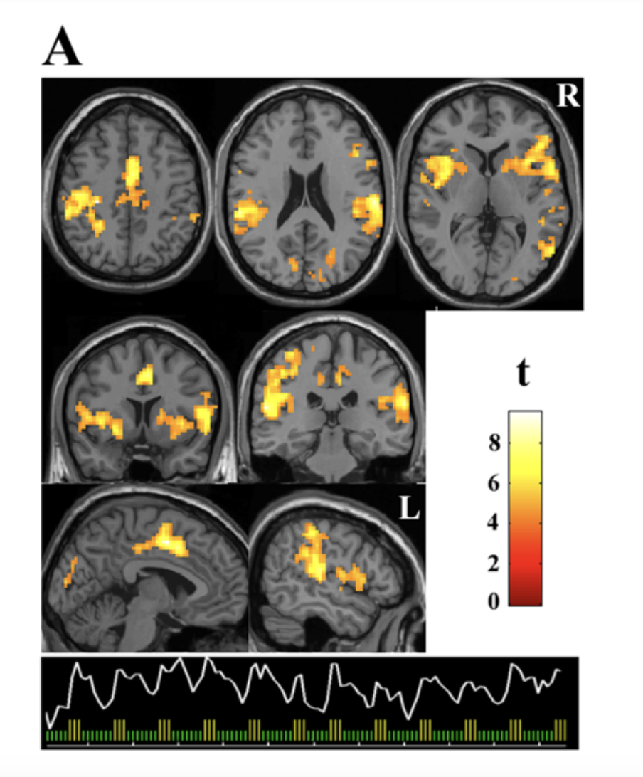
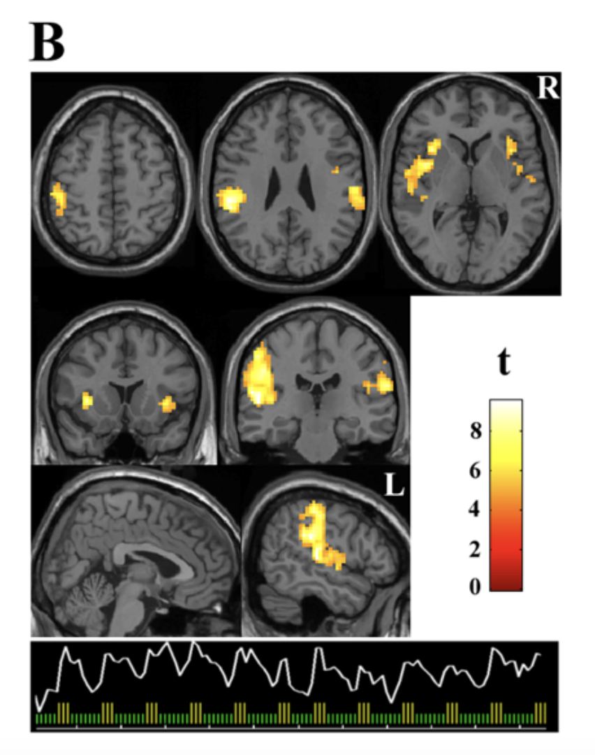
(t-value)
Fibromyalgia is a real, measurable condition associated with amplified pain processing in the brain.